
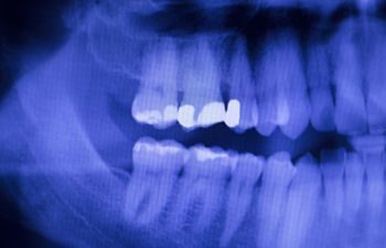
Low-dose imaging is used in all dental practices. These tools capture thorough images of your teeth and oral anatomy through the use of panoramic radiography to bitewings that focus on a specific area of your mouth.
Digital X-rays are used for clarity, guiding our dentist’s diagnostics to provide the best care for your family. When you have a better understanding of how X-rays work, you’ll see just how safe they truly are.
What are X-rays?
Dental X-rays are classified as Electromagnetic Radiation. These ray beams travel in a straight line in a wavelike motion that penetrates the X-ray films. You’re also exposed to radiation during everyday activities like spending a day at the beach or taking a flight in an airplane.
Any excessive radiation has the potential to cause, but dental radiography is extremely low dose. A full mouth series of films exposes our patients to less radiation than a transcontinental flight.
Today, dental machines contain strong filters and absorb unnecessary beams, while other parts in the machine limit the size and shape of the ray itself, reducing the amount penetrating specific tissue.
Dental practices measure for the maximum permissible dose (MPD) of radiation that a person can absorb with little or no injury. It is required of every staff member when providing for a patient. For instance, lead aprons with thyroid collars cover every patient during X-rays. This protects against any potential scattered radiation, even though low, from affecting reproductive organs.
Don’t Skip Your X-rays
Today, most dentists primarily use digital images that are even faster and use as much as 90% less radiation than traditional films.
All dental offices operate under proper radiation protocol for every individual, staff and patient alike. There is great peace of mind knowing that having dental X-rays taken is safe and healthy for your smile!